
What is FAI?
FAI stands for femoroacetabular impingement: a collision between the femoral head (femur) and the hip socket (acetabulum). This happens because one or both have a shape that differs slightly from average. During certain movements — particularly deep flexion and rotation — they collide slightly too early. This causes the labrum to become pinched and can damage it.
FAI is more common than you might think: a significant proportion of people have some form of cam morphology without experiencing any symptoms. We only speak of FAI syndrome when shape, symptoms and physical examination all point together towards impingement.
Two forms: cam and pincer
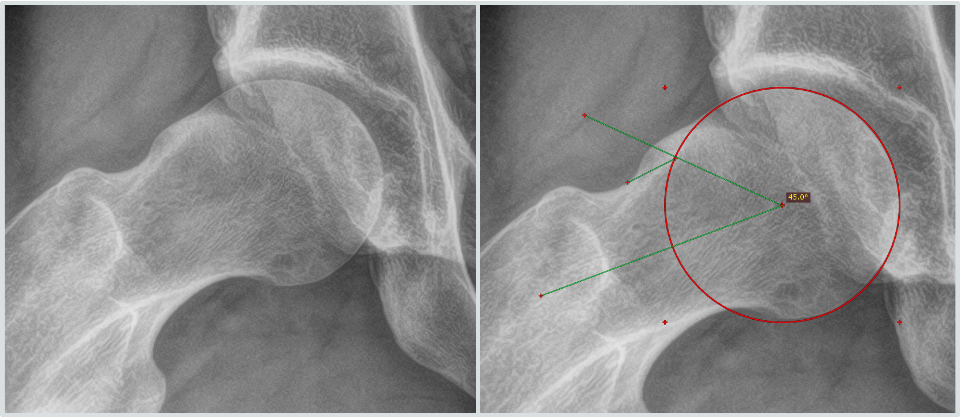
Cam impingement
With cam impingement, there is a bony thickening — which can be smaller or larger — at the junction between the femoral head and femoral neck. This bump collides with the labrum when you flex the hip, and can eventually also damage the adjacent cartilage. Cam impingement is more common in men, and more common in people who took part in intensive sport at a young age.
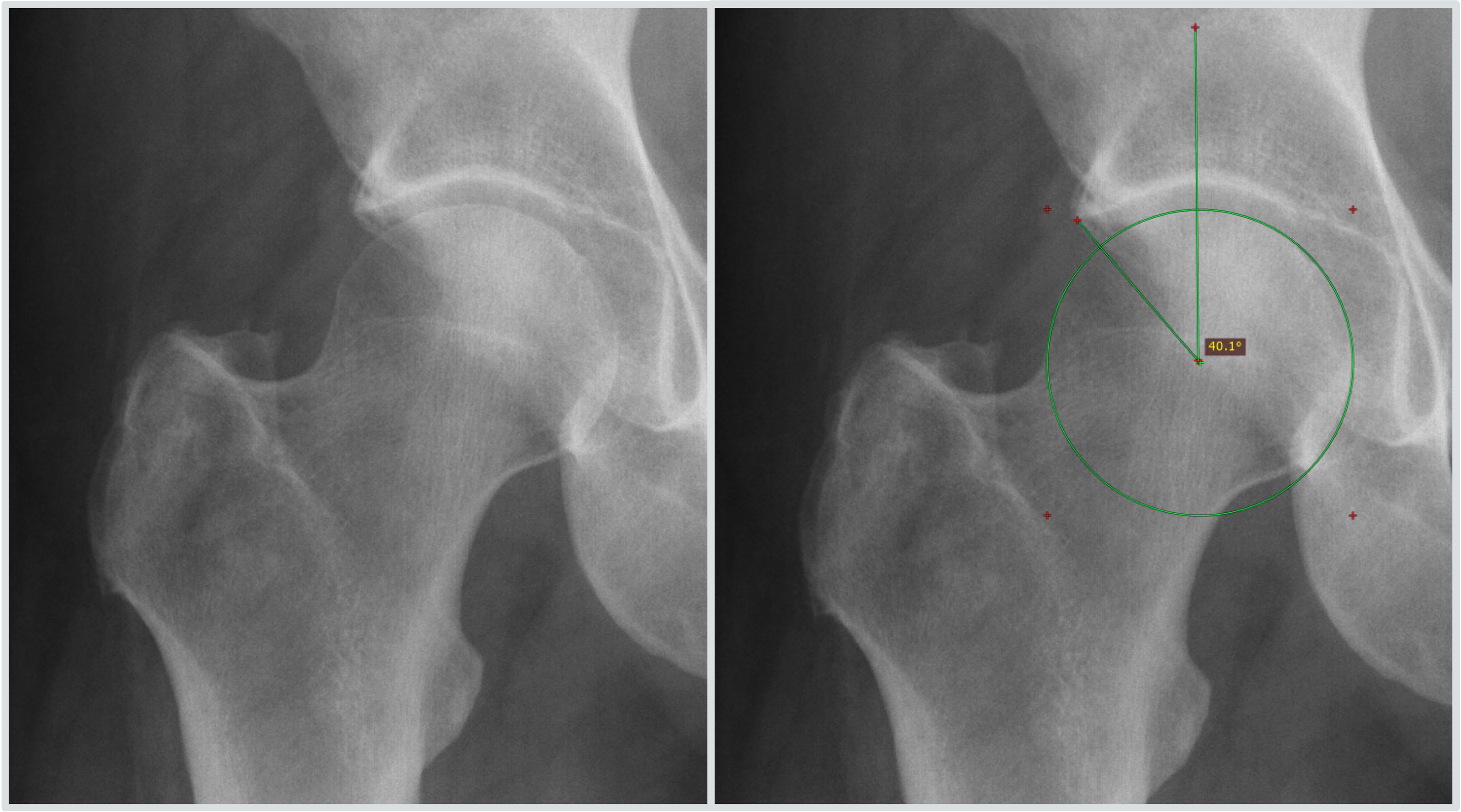
Pincer impingement
With pincer impingement, the rim of the hip socket covers the femoral head slightly too much. As a result, the femoral head collides with that rim earlier than it should. Pincer impingement is more common in women.
Mixed form
Many people with FAI have a combination of cam and pincer impingement.
Cam versus pincer at a glance
Location of the abnormality — cam: femoral head/femoral neck. Pincer: rim of the hip socket.
More common in — cam: men. Pincer: women.
Treatment during surgery — cam: shaving down the bony bump. Pincer: removing excess rim bone.
Risk of cartilage damage — cam: increased, particularly with longer-standing symptoms. Pincer: smaller, collides more with the labrum.
Symptoms
A characteristic feature of FAI is groin pain that occurs or worsens with movement: prolonged sitting, deep flexion (for example, tying your shoelaces), sport, or twisting movements. Reduced hip mobility, particularly on inward rotation, is also common.
Cartilage damage in FAI
With long-standing cam impingement, the bony bump collides with the cartilage at the rim of the hip socket with every flexion movement. Over time, this can lead to wear of that cartilage, often starting just next to the labrum. Because cartilage barely repairs itself, this is an important reason not to leave symptoms unaddressed for too long: in the longer term, particularly with the cam variant, this can contribute to the accelerated development of osteoarthritis (wear) of the hip joint. With pincer impingement, this risk is smaller: the collision mainly affects the labrum there, and the cartilage itself to a lesser extent.
How do we diagnose FAI?
The diagnosis of FAI syndrome is made on the basis of three things together: your symptoms, the findings on physical examination — such as a test in which the hip is flexed, rotated inward and brought towards the abdomen — and the shape of your hip on an X-ray or MRI scan. Read more about these examinations on the page about imaging investigations. An abnormal shape on a scan alone is therefore not enough for the diagnosis: many people with that shape never have any symptoms.
Treatment
Exercise therapy is the first step for FAI and helps a proportion of people sufficiently; your physiotherapist will also look with you at whether activities at work, in sport or in hobbies can be temporarily adjusted. If this does not give sufficient results, a hip arthroscopy may be considered: this involves shaving down the colliding bony overgrowth (cam) or removing the excess rim bone (pincer), repairing a damaged labrum with stitches — or, if that is no longer possible, removing the damaged part — and treating any cartilage damage during the same procedure. Read more about weighing up exercise therapy against surgery on the page about alternatives.