
X-rays: seeing the shape of your hip
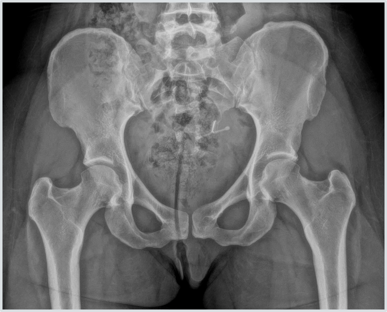
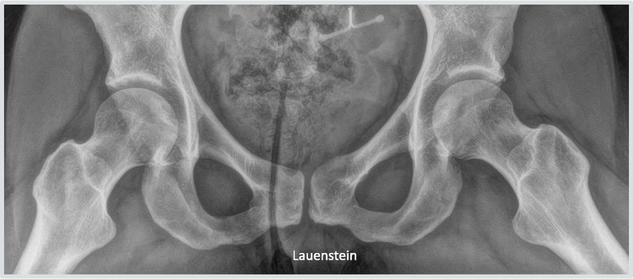
An X-ray of your pelvis is often the first step. It clearly shows the shape of your femoral head and hip socket. Usually two images are taken: an AP pelvis X-ray and a Lauenstein X-ray.
AP pelvis X-ray
This image of your whole pelvis, taken straight from the front, shows how much the rim of your hip socket covers the femoral head — too much coverage is consistent with pincer impingement. This image also shows whether the hip socket sits in a normal position or is slightly rotated (retroversion).
Lauenstein X-ray
For this image, you lie with your hip in a specific, slightly bent and rotated position (a 'frog-leg position'). This best shows the junction between the femoral head and femoral neck — the site where a cam lesion develops.
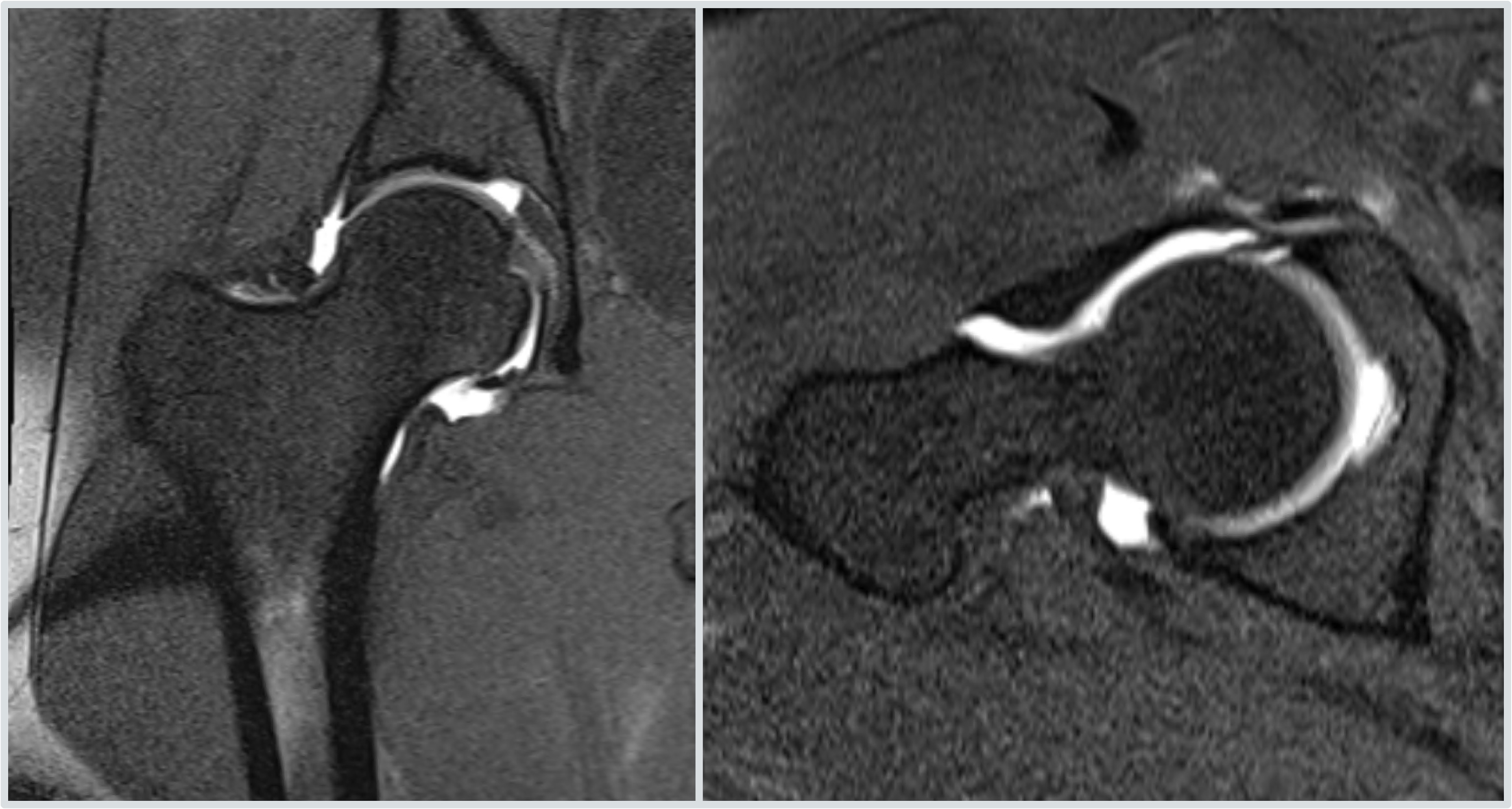
MRI: seeing the labrum and cartilage
An X-ray mainly shows bone. A clear picture of the labrum and cartilage requires an MRI. For this, an injection is often first given into the hip joint under ultrasound guidance, delivering a contrast fluid (an MRI arthrogram). This contrast spreads through the joint and makes the labrum and any cartilage damage considerably clearer on the MRI images taken afterwards.
Diagnostic injection (marcaine test)
Sometimes it isn't immediately clear whether your hip joint itself is the source of your symptoms. In that case, a diagnostic injection under ultrasound guidance can help — also known as a marcaine test. For this, your orthopaedic surgeon injects a combination of a local anaesthetic (lidocaine) and an anti-inflammatory (corticosteroid) into your hip joint.
If your pain clearly eases in the hours afterwards, this is a strong indication that the hip itself is the source of your symptoms. After the injection, you'll keep a brief diary: what differences do you notice in the first hours, the first days and the first weeks, compared with the period before the injection? After two to three weeks, we discuss together — often by phone — what the effect has been.
Rarely needed: CT or bone scan
In a small number of cases, a CT scan or bone scan provides additional useful information, for example about the precise shape of the bone or the extent of osteoarthritis. We only use this investigation when it's genuinely necessary.